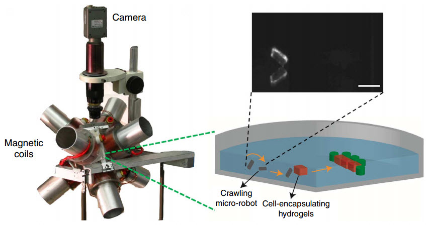
 A new breed of micro-robots has been demonstrated to be capable of constructing complex 3D printed tissue architecture by gently guiding diverse cell-encapsulating building blocks, known as hydrogels, to their proper places in multi-layered and heterogeneous tissue structures.
Developed by researchers at Brigham and Women’s Hospital and Carnegie Mellon University, this pioneering technique combines recent advancements in tissue engineering, 3D printing and microrobotics. The scientists used magnetic fields to remotely enable these tiny robots – fashioned as a sort of microscale tweezers – to move one cell at a time, change its orientation and place it where needed with a precision and at a scale we previously thought to be unimaginable.
The study, published in Nature Communications, suggests that, unlike other existing bioprinting methods, this technique allows for precise modification of tissue architecture and is easily reversible. In other words, if before a misplacement of a cell-containing droplet could ruin the process in a blink of an eye, now a micro-robot can remove the misplaced droplet and place it where it belongs. This is the first time that cells are manipulated one by one in a 3D printing process with a full control of time and position.
Bioprinting and tissue engineering, for some time now, have been pushing the boundaries of our imagination by promising to bring about the possibility of creating new donor organs as well as devising new therapies and testing drugs using way more practical cell-based models. Thanks to micro-robots this possibility has just become one step closer.


